08 9393 3373
08 9393 3373
08 9393 3373
VETERINARY HOSPITAL
08 9393 3373
West Coast
West Coast Veterinary Hospital offers a full range of equine services. Need to find out more. Please get in contact with our team for any of the following services.


FEATUREd Services
Once bookings are made our reception team will be in contact regarding an appointment time!

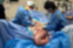
Gastroscope
Concerned if your horse has ulcers? Performing a gastroscope is the simply way to give you an accurate diagnosis.

DYNAMIC SCOPE
West Coast Veterinary Hospital are proud to add another diagnostic modality alongside the Nuclear Scintigraphy suite, MRI, Radiography and Ultrasonography. The Dynamic Equine Exercise Scope is a game changer for analysing upper airway issues in equine athletes when in work. Airway issues are common in sporthorses and racing horses and can effect performance and general health.
Dynamic Scopes are $550

Basic Equine Dental, Randlab Oral Wormer and Electrolyte DrenchchCC
Dental / Drech and worm

Shockwave Therapy is an extremely useful treatment for soft tissue injuries. It can aid in healing of tendon and ligament injury and may even help treat pain associated with kissing spines. Many bowed tendons or suspensory ligament injuries will benefit from such treatment.
Save 20% and book 4 treatments for $425
shockwave
Interested in finding out more about common equine related issues? Our team have helped cover some main important aspects of equine conditions.
It is always an alarming sight when discovering a horse with a sore eye. In most cases, with early intervention, ophthalmic (eye) cases can be managed successfully without major complications. However, in some instances, a horse with an eye problem is a veterinary emergency. Merely waiting 24 hours without medical intervention can result in loss of the eye. The crux of the matter is being able to differentiate the simple from the complex cases. Unless an owner is confident it is always best to seek medical intervention early.
Blocked tear duct: Excessive tear production and conjunctivitis are seen in all ages of horses. Blocked tear ducts can be managed effectively by 'flushing'. This is when a small tube is placed up the nasolacriminal duct (connection between the eye and nasal passage) and water is used to irrigate the canal. Some horses require regular flushing whilst others will clear after just one treatment. Occasionally this blockage of the tear duct is due to a low-grade chronic infection and the animal will require medication directly into the eye for several days.
Cornea damage: After digital palpation to determine if there are any foreign bodies within the eye (e.g a grass seed) the corner is examined for damage to the cornea will stain bright green or orange. Very deep ulcers, down to the deepest layer of the cornea (descents membrane), will not stain. This condition is called a 'descemetocele'. This is a serious condition that requires immediate medical attention. If this condition is eft untreated the globe (eyeball) may rupture and the eye may need to be removed. Medications with cortisone-based products should be used with cation in the equine eye. this is because, if an ulcer is present, healing may be severely impaired and secondary viral infections are even possible. An eye should always be stained with fluorescent prior to steroid based medications being used. A common complaint is that a horse will become very intolerant to receiving medications into the eye. Sub-palpebral lavage systems are used in such situations. Medications can be injected from a remote site. (e.g near the neck) and a direct line that is situated under an eyelid will dispense the medications into the eye.


Equine Ophthalmology:
Cancer of the third eyelid:
A condition that is frequently seen in draft breeds and grey/pale skinned horses is cancer of the third eyelid. The third eyelid, also known as nictitans membrane, is a structure that will flick from the inside to the outside aspect of the year in order to spread the tear film. The condition is usually caused by squamous cell carcinoma, a locally invasive tumor caused by sun exposure that can spread if not treated. this condition requires surgical removal of the majority of the third eyelid. Removal of this eyelid is tolerated extremely well and will almost always result in a cure of the condition and have no lasting negative effects.
Eye Removal:
In the worst-case scenario, when the eye cannot be saved or if radical surgical intervention is prohibited by economics, the eye must be removed. horses tolerate this procedure extremely well and in most cases it can be performed standing under local anesthetic. Some owners are alarmed at the final cosmetic result, however the horse never seems to be bothered. It is sometimes possible to insert prosthetic globes under the remaining skin in order to give the appearance of a normal eye. It may not be the most aesthetically appealing outcome but most athletic disciplines can still be performed successfully even if the horse has one eye (or vision in one). Some horses more easily spook and depth perception may be altered therefore caution is advised with competing in some sports. It should also be noted that with racing Thoroughbreds, the rules of racing state that the animal must have vision in both eyes.
Any eye issue is considered an emergency and should be dealt with as soon as possible. If you have concerns your horse may have any of the above we urge you to make an appointment on Ph: 9393 3373
Nasal and Sinus Issues
Sinusitis: Excessive tear production and conjunctivitis are seen in all ages of horses. Blocked tear ducts can be managed effectively by 'flushing'. This is when a small tube is placed up the nasolacriminal duct (connection between the eye and nasal passage) and water is used to irrigate the canal. Some horses require regular flushing whilst others will clear after just one treatment. Occasionally this blockage of the tear duct is due to a low-grade chronic infection and the animal will require medication directly into the eye for several days.
Ethmoid hematoma: After digital palpation to determine if there are any foreign bodies within the eye (e.g a grass seed) the corner is examined for damage to the cornea will stain bright green or orange. Very deep ulcers, down to the deepest layer of the cornea (descents membrane), will not stain. This condition is called a 'descemetocele'. This is a serious condition that requires immediate medical attention. If this condition is eft untreated the globe (eyeball) may rupture and the eye may need to be removed. Medications with cortisone-based products should be used with cation in the equine eye. this is because, if an ulcer is present, healing may be severely impaired and secondary viral infections are even possible. An eye should always be stained with fluorescent prior to steroid based medications being used. A common complaint is that a horse will become very intolerant to receiving medications into the eye. Sub-palpebral lavage systems are used in such situations. Medications can be injected from a remote site. (e.g near the neck) and a direct line that is situated under an eyelid will dispense the medications into the eye.
Wounds and fractures: A condition that is frequently seen in draft breeds and grey/pale skinned horses is cancer of the third eyelid. The third eyelid, also known as nictitans membrane, is a structure that will flick from the inside to the outside aspect of the year in order to spread the tear film. The condition is usually caused by squamous cell carcinoma, a locally invasive tumor caused by sun exposure that can spread if not treated. this condition requires surgical removal of the majority of the third eyelid. Removal of this eyelid is tolerated extremely well and will almost always result in a cure of the condition and have no lasting negative effects.

Sinus cysts: In the worst-case scenario, when the eye cannot be saved or if radical surgical intervention is prohibited by economics, the eye must be removed. horses tolerate this procedure extremely well and in most cases it can be performed standing under local anesthetic. Some owners are alarmed at the final cosmetic result, however the horse never seems to be bothered. It is sometimes possible to insert prosthetic globes under the remaining skin in order to give the appearance of a normal eye. It may not be the most aesthetically appealing outcome but most athletic disciplines can still be performed successfully even if the horse has one eye (or vision in one). Some horses more easily spook and depth perception may be altered therefore caution is advised with competing in some sports. It should also be noted that with racing Thoroughbreds, the rules of racing state that the animal must have vision in both eyes.
The Equine Mouth
Common teeth issues: Excessive tear production and conjunctivitis are seen in all ages of horses. Blocked tear ducts can be managed effectively by 'flushing'. This is when a small tube is placed up the nasolacriminal duct (connection between the eye and nasal passage) and water is used to irrigate the canal. Some horses require regular flushing whilst others will clear after just one treatment. Occasionally this blockage of the tear duct is due to a low-grade chronic infection and the animal will require medication directly into the eye for several days.
Choke: After digital palpation to determine if there are any foreign bodies within the eye (e.g a grass seed) the corner is examined for damage to the cornea will stain bright green or orange. Very deep ulcers, down to the deepest layer of the cornea (descents membrane), will not stain. This condition is called a 'descemetocele'. This is a serious condition that requires immediate medical attention. If this condition is eft untreated the globe (eyeball) may rupture and the eye may need to be removed. Medications with cortisone-based products should be used with cation in the equine eye. this is because, if an ulcer is present, healing may be severely impaired and secondary viral infections are even possible.
Jaw and dental fractures: A condition that is frequently seen in draft breeds and grey/pale skinned horses is cancer of the third eyelid. The third eyelid, also known as nictitans membrane, is a structure that will flick from the inside to the outside aspect of the year in order to spread the tear film. The condition is usually caused by squamous cell carcinoma, a locally invasive tumor caused by sun exposure that can spread if not treated. this condition requires surgical removal of the majority of the third eyelid. Removal of this eyelid is tolerated extremely well and will almost always result in a cure of the condition and have no lasting negative effects.

Cyst: Excessive tear production and conjunctivitis are seen in all ages of horses. Blocked tear ducts can be managed effectively by 'flushing'. This is when a small tube is placed up the nasolacriminal duct (connection between the eye and nasal passage) and water is used to irrigate the canal. Some horses require regular flushing whilst others will clear after just one treatment.
Occasionally this blockage of the tear duct is due to a low-grade chronic infection and the animal will require medication directly into the eye for several days.
The Equine Ear

Keyhole Surgery
Laparoscopy: Laparoscopy is when a camera is placed into the abdomen of the horse - which has been distended with carbon dioxide - to visualise the internal organs. The laparoscopy is attached to a video camera which displays the image on a monitor screen. The entire procedure is done under sedation and local anaesthetic. Only small, 1-2cm incisions are required in most circumstances. Laparoscopic procedures in horses are less invasive and are associated with less postoperative pain and inflammation. One of the latest advancements with 'key hole' surgery in the equine field is the use of laparoscopy to treat barren or sub-fertile mares. Debris within, or blocked oviducts, (the pipe that allows the egg to travel from the ovary to the uterus) are responsible for many failed pregnancies. Problem with the oviducts are usually a diagnosis reached after all other possible causes of infertility have been excluded. The application of PGE2 gel onto the surface of the oviducts is the treatment of choice for oviduct blockages. This medication will help the oviducts clear debris or blockages. The laparoscopy is done with the horse is standing in stocks, via laparoscopy. Many recent studies have shown that blocked oviducts are responsible for failed pregnancies of barren mares. Success rates (pregnancies) can be up to 90% of horses following application onto PGE2 gels on oviducts. Many of these horses treated are in their late teens and may have been barren for several years. Mares can be inseminated during the same cycle as the procedure and, due to the procedure being 'key hole', they recover within days. Mares that obtain a pregnancy following the procedure are excellent candidates for the same treatment the following year.
Laser Surgery: Lasers have made their way into the veterinary field. From sarcoids to upper respiratory tract surgery - they have many uses. The removal of vocal cords, ventricles and treatment of soft palate problems are all possible. With the aid of local anaesthetic and sedation the laser fibre can be passed up an endoscope portal, which has been placed into the upper respiratory tract via the horse's nasal passage. The area of concern is then grasped with the aid of long forceps and the laser can be aimed to cut the problematic tissue. The surgical field is visualised on a monitor next to the standing animal.
Dental Procedures: The extraction of upper cheek teeth can be a complicated ordeal at the best of times. Many horses require their teeth surgically removed and or repelled from the sinuses. This can be very traumatic and complications often occur. A new technique is now available that is minimally invasive transbuccal approach, which allows extraction of upper cheek teeth. This is ideal for teeth that cannot be removed easily with molar extractors via an oral approach (loose teeth). A small incision (1cm) is made into the cheek and a hole is drilled into the tooth under endoscopic visualisation. An anchor is then screwed into the tooth which is then removed via repulsion. Owners should consider this if upper cheek teeth require removal.


Tenoscopic Surgery: The most commonly accessed tendon sheath is the digital flexor tendon sheath. This runs down the back of the fetlock to the pastern in all four limbs. It contains the flexor tendons, annular ligaments and other important soft tissue structures. Externally these swellings appear as ' windgalls; which are rarely a problem. However, in some instances (lameness, infection or severe swelling) they require surgical treatment. A camera is placed into the sheath after it has been distended with sterile fluid. Small incisions then allow a camera to be placed into the sheath via small 1cm portals. this 'key hole' surgery technique excellent visualisation of all the structures within the sheath and allows accurate treatment. Horses recover quickly and only need a few weeks box rest.
Arthoscopy Surgery: The joints that can be accessed via arthroscopy include; coffin, pastern, fetlock, carpal, elbow, shoulder, hock, stifle, hip, mandibular and even articular facet joints within the neck. Arthroscopy is when a small camera is placed into a joint and 'key hole' surgery us performed to treat problem joints. Previously, a large incision into the joints would have been required, which led to long healing times and permanent lameness. Nowadays with the use of arthroscopic cameras the prognosis for many conditions is excellent. Conditions that are commonly treated with arthroscopy include the removal of bone fragments e.g OCD fragments) or treatment of damaged soft tissue structures within joints (cartilage, ligaments and inflamed synovium). Arthroscopic surgery can now be performed standing in some circumstances.
Ulcers in horses can cause discomfort, poor performance, loss of appetite, failure to thrive, a poor coat and even colic or internal bleeding. Although it is believed that stress, exercise, poor intermittent access to feed and confinement can factor EGUS in horses; there is not one single cause. These can all lead to the production of excessive levels of gastric acid which then erodes the wall of the stomach to cause ulcers.
Ulcers: It's what you can't see. The signs of ulcers are often hard to detect as they happen inside to stomach and can be confused with other conditions. Ulcers are formed when the mucosal lining of the stomach is eroded. The best way to see if your horse has ulcers is to perform a gastroscope. This allows your veterinarian to see inside the horses stomach and determine the grade of ulcers and then recommend the best treatment for your horse.
Horse’s are likely to stress when either travelling to or once at a competition. Here are 10 tips to help relieve stress in your horse when travelling.
If you are taking your horse on a long trip, the level and severity of the stress will be higher and may have serious consequences. Stress lowers a horse’s resistance to disease, and those stressed by transport are vulnerable to gastric ulcers, pneumonia, diarrhoea, colic or laminitis. Transport stress the sum of adverse effects on the horse during transportation. There are a lot of contributing factors – discomfort in a particular trailer, noise, road conditions, confinement. Also footing and balance issues, changes in feeding and watering, temperature and humidity. Separation from herd mates, new environments, or interactions with unfamiliar horses can impact stress levels. Being aware of the causes means you can take steps to minimise them. That way you can reduce the impact of transport stress and arrive at your destination with a happier, healthier horse.
-
Take a Break: Every four hours, give your horse a chance to urinate, and walk around a bit. Comfort breaks take a bit of pre-planning, but the reduction in fatigue and stress to your horse is well worth the effort.
-
Horse Hydration: It's easy for horses to become dehydrated during transportation. Offer them water periodically. Adding an electrolyte will help rehydration and give them energy for the journey.
-
Feed Well: Offering travelling horses their regular feed helps them stay calm, retain water in their gut, and reduces the risk of gastric ulcers and colic.
-
Lung Health: Pathogens and dust from hay and dried manure can quickly enter a horse's respiratory system. Wetting hay can help, and always make sure the trailer is cleaned thoroughly before any trip. If there is a history of inflammatory airway disease, consider therapy before travelling.
-
Let the Air in There: Proper ventilation keeps your horse cooler and less susceptible to potentially toxic airborne particles in the trailer.
-
Head Down: Allowing them the ability to lower their heads below their shoulders means horses are less likely to suffer respiratory stress. It's how they naturally carry their heads, allowing them to breathe comfortably. Keep in Mind Who's Behind: Remember, a slow and steady journey is not only safer, but it's also much less stressful for the horse.
-
Learn to Load: Loading is often the most stressful part of the trailering experience for horses. It's a good idea for you to practice loading your horse several times a year, so you're both comfortable with the procedure.
-
Monitor Vital Signs: Learn how to measure your horse's vital signs during travel. If your horse does get sick or shows signs of pain during the trip, you'll be able to give the vet the information they need.
-
Be Prepared: Always carry an emergency first aid kit, including an extra supply of your horse’s medications. Learn how to recognise the signs of transport stress such as dehydration, ulcers and colic. And carry extra feed supplies. You never know, your trip may take longer than planned.
Gastric Ulcers

Lameness Examination:
Being able to tell if a horse is lame and determining where the problem originates can be difficult at times for even the experienced horse person. Not all lamenesses appear as a horse ‘limping’, some may just be a bobbing of the head, a change in the way the horse carries itself overall, or just a change in attitude. Many riders can ‘feel’ something is just not right with their horse, often before there is visually a noticeable difference in movement, and early intervention by a veterinarian can often prevent further damage.
To be able to identify and understand lameness in horses, which can involve virtually any part of the body and can originate in bone or soft tissue, riders and owners must know how to distinguish between normal and altered movement. Lameness is defined simply as an abnormality in a horse’s movement caused by pain or reduced range of motion (see image 1) and the term covers a wide range of ailments. A horse that is lame is often referred to as being unsound.
The most common health problem seen in the equine athlete; as a general rule 75% of lameness issues are seen in the forelimb whilst the remainder are hind limb issues. Of the lameness problems seen in the forelimb 95% of these originate from below the knee or hock. Some horse owners think of lameness only as a problem of the feet or legs and never look further than these areas, however, the most commonly misdiagnosed lameness issues in horse are suspected shoulder and hip lamenesses. It is always a wise idea to start by examining the hoof initially and then looking for other explanations.
Locating the source of lameness and then developing a plan to manage it are high priorities and these procedures will generally involve the veterinarian or farrier, or often both.
How to determine if a horse is lame: A lame horse is in pain. As an owner it is your responsibility to be able to tell if the horse is lame and then seek appropriate treatment. When veterinarians are assessing lameness, horses are commonly graded from 0-5 based on the level of the lameness. This scale indicates the severity of the lameness. This 0-5 scale had originated from the AAEP (American association of Equine Practitioners) system and is as follows:
Lameness not perceptible under any circumstances.
Lameness is difficult to observe and is not consistently apparent, regardless of circumstances.
Lameness is difficult to observe at a walk or when trotting in a straight line, but consistently apparent under certain circumstances (e.g., weight carrying, circling, inclines, hard surfaces, etc.)
Lameness is consistently observable at a trot under all circumstances
Lameness is obvious at a walk
Lameness produces minimal weight bearing in motion and/or at rest or a complete inability to move.
Terms such as ‘acute’ and ‘chronic’ are sometimes used when assessing lameness. Both terms are used to explain the duration of the condition. Acute means that the condition has occurred suddenly and has not been present for a long period of time. Chronic conditions are of long-standing duration that may have been present for several weeks to months. The severity of the lameness (see above) is an independent scale to that of the duration.
Other important variables: Other variables of lameness are whether it is persistent or intermittent, and progressive or static. There is uncertainty amongst horse owners with these terms, some confusing persistent with chronic, but chronic refers to the length of time of the problem while persistent means that the lameness (which could be recent or not) has been consistently observable since its onset – not coming and going (e.g. intermittent). Unless the horse is non-weight bearing (5/5 lame) or lame at the walk (4/5 lame) lameness is most easily assed while the horse is trotting. To fully recognise the extent of lameness the horse should be observed at the trot in a straight line and on circles both ways while on the lunge. This should be done with the horse being worked on both a hard and soft surface that is even and free of debris, as an area strewn with debris is not going to give a true indication of the horse’s lameness.
Many high-speed lameness problems are only present when the horse is cantered therefore the horse should also be viewed in this gait if appropriate. If the pain is only in one leg it should be easy to notice that the horse is not moving evenly. Some horses can be lame in multiple limbs – which makes diagnosis much more difficult. A sound horse should move evenly in its stride with minimal head movement. The hips should rock evenly behind with no toe drag along the ground. (see image 3) A lame horse will often throw its head in rhythm with its stride. If the horse is sore in a front leg, it will throw its head up as the sore side touches the ground or down when the sound leg hits the ground (see image 4 ). ‘Head nodding’ is typically associated with forelimb lameness.
If the lameness is in a back leg, the horse will lean (the hip) onto the sound side (down on sound). He may also drag the toe of the lame limb. (see image 5). Hip drop. If the horse is painful in both front feet (or hind) it will typically move with a very shuffled gait and only be obviously lame when lunged in either direction.
Once it has been determined that the horse is actually lame, what is the next step? Always check the foot! Hoof abscesses are also known as ‘the great pretender’ and due to the level of pain (severe) being displayed by the animal they are commonly mistaken as a fracture. While more common in the front feet they do occur in hind hooves.
Depending on the level of lameness the veterinarian and or farrier will conduct an initial examination that would include an accurate medical history, full physical examination and a more specific lameness examination. If your veterinarian has been called out it is likely he or she will palpate (see image 7 ) the affected limb and even perform flexion tests to try and isolate the lameness. Things they will be feeling for are increased heat, swelling and pain of specific areas. Flexion tests are when an area of the horse’s limb is held in ’flexion‘ for a set amount of time and then the horse is trotted off. A positive response is when the horse becomes significantly lamer after the test.. (see image 9 )
In these illusive cases nerve blocks can be performed. Nerve blocks (see image 10) are when the nerve or nerves to a specific anatomical location are desensitised by infusing local anesthetic into the area. They can be performed on most areas of the front and hind limb and can be extremely helpful in identifying the area of pain. A horse will typically become sound once local anesthetic has been applied to the location causing pain (e.g. the fetlock joint). The appropriate diagnostic modality (e.g. ultrasound or radiography) can then be used to focus on and further assess the area.
Occasionally lamenesses are not a leg issues and the pain is originating from anywhere from the back (e.g kissing spines – see image 11), head, neck, or pelvis. These cases can be very complex and apart from nerve blocks some other more advanced diagnostics tests are required. Nuclear scintigraphy (aka ‘bone scanning’) can be used to assess structures such as the neck and pelvis more easily. Lamenesses can become extremely complex and occasionally involve multiple limbs, however this article is aimed at giving a basic understanding on how lameness can be detected and then localised to a specific area. Involving a veterinarian early once a lameness is noticed can significant improve the chances of the horse becoming sound, as appropriate treatment can be undertaken promptly.
Common causes of fore limb lameness:
Foot - Solar bruising, hoof abscess, poor shoeing, greasy heel, laminitis
Fetlock - Trauma, soft tissue damage, osteoarthritis, osteochondral fragment (bone chip)
Knee - Osteoarthritis, osteochondral fragment (bone chip)
Shoulder - OCD, muscular injury
Common Causes of hind limb lameness:
Foot and Fetlock - Refer to forelimbs
Hock - OCD, lower joint arthritis (bone spavin)
Spine - Muscular pain, kissing spines, poor saddle fit
Advanced Equine Imaging: Our hospital is proud to offer Western Australia's only veterinary nuclear scintigraphy and MRI service a long with radiography and ultrasound.
Nuclear Scintigraphy - also known as 'bone scanning' is a process used in horses to diagnose lamenesses. Nuclear Scintigraphy is the only way to reliably investigate upper limb lameness, horse's that cannot be nerved blocked or have complex multiple limb lameness. It is extremely safe with no general anaesthesia required.
MRI - Stationary MRI is the standard of excellence recognised worldwide as the correct way to scan anatomy without compromising image quality. The o-scan equine covers imaging of the foot, pastern, fetlock, high suspensory, carpus and tarsus regions of the equine limb. The o-scan equine is the ideal MRI to clearly distinguish between bone edema, ligaments and tendons ensuring accurate imaging, through excellent contrast resolution and image quality; essential for optimal diagnosis.


Equine MRI
The science of equine MRI is growing rapidly, and knowing what to do with the information gained from an MRI study is the key to a sound horse. MRI or Magnetic Resonance Imaging is an imaging modality that utilizes the magnetic forces within the patientís cells to create an image. This allows both bone and soft tissue structures to be seen with excellent detail. Additionally, information on the underlying disease process can often be obtained from the MR image. No other diagnostic tool gives more detailed information regarding pathology in the anatomic structures involved. It eliminates questions and provides answers that no other diagnostic tool can provide.
MRI technology is the ìgold standardî in medical imaging of soft tissues including tendon, ligament, joint capsule and articular cartilage, giving exquisite anatomical detail and precise localisation of lesions. MRI also provides precise localisation and differentiation of various pathological processes in bone. MRI finds its greatest use in lameness conditions of the lower limb, principally the foot and has revolutionised the diagnosis of injuries/conditions formerly collectively grouped as ìnavicular syndromeî.
Nuclear Scintigraphy
The Equine Hoof:
The equine hoof is a complex structure surrounding the distal phalanx of the 3rd digit of each of the four limbs of which is covered by complex soft tissue and keratinised structures. With its complex structure comes many common issues;
Laminitis: Laminitis is a crippling disorder of the feet, resulting from damage to the sensitive soft tissues known as 'laminae' which connect the skeletal pedal bone to the inside of the hoof wall. There are about 600 interlocking laminae in each hoof and they provide the support for the weight of the horse. Founder is the physical change that occurs within the hoof as a result of laminitis. When the laminae are damaged their supporting function is weakened. This may allow the pedal bone within the hoof to 'sink' downwards and 'rotate' backwards due to the weight of the horse pushing downwards on the bone and the upward pull of the flexor tendon which attaches to the base of the bone. Damage to this bone can lead to abnormal foot growth with obvious 'growth rings', long toes, flat soles and separation of the sole from the hoof wall at the white line (often followed by seedy toe).
Thrush and Fungal Infections: Fungal infections in the hoof are common in the winter months. Bacteria and fungi cause hoof disease which harms sensitive hoof tissue. It is important to keep the feet dry and clean, either by cleaning with a iodine solution or using specific trush and bacteria fighting remedies West Coast Vets stock the following products to help you fight thrush and fungal disease in your horse.
SBS Thrush Stop
Hawthornes Sole Pack


Castration and Cryptorchids (Rigs):
Castration (gelding) has been used for hundreds of years to control unwanted aggressive behaviour along with taking away the ability to reproduce. It is one of the most common surgeries performed by the equine practitioner. The testicles are the organs responsible for producing testosterone, the main hormone that produces the stallion-like behaviour and normally these descend from the abdominal cavity into the scrotum just before or after birth. There is no ideal 'ideal' time to castrate a horse. However most people castrate their horses as yearlings, before they can develop these stallions-like mannerisms.
Castration: If both testicles have descended normal it is routine surgical procedure to castrate the animal. On an interesting note the age of when you castrate a horse will influence how tall they grow. Horses castrated young (younger than 6 months of age ) tend to grow tall. This is because the male hormones produced by the testicles tend to increase muscle mass and decrease vertical growth.
Cryptorchids (Rigs): However one or both testicles fail to descend into the scrotal sac. Removing one testicle is not ideal as this will not stop the stallion-like bahaviour occurring because the internal testis continue to produce testosterone and male hormones regardless of where it is in the body. The undescended testicle can be located anywhere from the inguinal ring/canal 'high flanker' to completely within the abdomen. The majority of left hand side testicles are retained within the abdomen whilst right testicles are commonly 'high flankers'. Most common is one testicle affected however bilateral is possible. It is important to know if the horse is a cryptorchid not retractile. A retractile horse is one that ' retracts ' the testicle up towards the abdomen when palpated (handled). Removal of a cryptorchid testicle is much more difficult than removing testicles that have descended. Traditionally these have been resolved under general anaesthetic with the horse on its back. Complications such as muscle or nerve paralysis, along with limb fracture can occur the horses recovering from a general anaesthetic. Laparoscopic castration, the use of small surgical portal into the side of the abdomen with the horse standing is a new technique for treating cryptorchidism.This method requires the horse sedated and local anaesthetic injected into the skin. The laparoscope is inserted into the side of the abdomen and the internal testicles grasped. Prior to insertion of the laparoscope the abdomen is inflated with carbon dioxide to aid with visualisation and locating the internal testicle. The testicle is then visualised and removed either with the aid of a ligating devise. The small portal holes are then closed with sutures and the horse kept in hospital for 24 hours. Very low complications rates are associated with this surgery technique.

